Case Identification
Case ID Number
Tumor Type
Body region
Position within the bone
Periosteal reaction
Benign or Malignant
Clinical case information
Case presentation
This 18 year old male has pain in the shin for 4 months. The pain is exacerbated by his ongoing training for the "iron man" competition. Examination of the tibia shows there slight point tenderness, but no mass or swelling. No regional lymphadenopathy. Knee exam is normal.
Radiological findings:
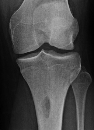
Plain radiographs show a well-circumscribed proximal tibial lesion, with a sclerotic rim. The lesion has slightly expanded the anterior cortex of the tibia. It extends through the anterior cortex into the medullary cavity. It is radiolucent, there is no matrix mineralization. It appears that there is diffuse mineralization or increased density of the bone surrounding the lesion for several centimeters proximal and distal which has a very even non-flocculent appearance. Technetium 99 bone scan shows that there is intense abnormal uptake in the area of the lesion and no other skeletal lesions are seen.
An MRI shows that the lesion has a well-circumscribed sclerotic rim, it measures approximately 4 cm from proximal to distal, and about 3 cm from anterior to posterior.It has slightly expanded the cortex and reaches from outside the tibia on the anterior medial surface across the full width of the cortex into the central medullary canal, at which point it is well bounded by a relatively dense sclerotic rim. On one sagittal view, there is a faintly seen dense horizontal line at the level of the lesion and in the center of the reactive zone around the lesion, which would be characteristic of a stress fracture. Within the lesion itself, there are multiple lobular areas that have variable signal intensity, with some very bright, some intermediate, and there are what appear to be internal septations. Surrounding the lesion, and extending proximally 6 to 8 cm all the way across the location of the proximal tibial physis, and extending distal from the lesion for 4 to 6 cm is a diffuse increase in bone marrow signal intensity, suggesting reactive change. There is no definite periosteal reaction.
An MRI shows that the lesion has a well-circumscribed sclerotic rim, it measures approximately 4 cm from proximal to distal, and about 3 cm from anterior to posterior.It has slightly expanded the cortex and reaches from outside the tibia on the anterior medial surface across the full width of the cortex into the central medullary canal, at which point it is well bounded by a relatively dense sclerotic rim. On one sagittal view, there is a faintly seen dense horizontal line at the level of the lesion and in the center of the reactive zone around the lesion, which would be characteristic of a stress fracture. Within the lesion itself, there are multiple lobular areas that have variable signal intensity, with some very bright, some intermediate, and there are what appear to be internal septations. Surrounding the lesion, and extending proximally 6 to 8 cm all the way across the location of the proximal tibial physis, and extending distal from the lesion for 4 to 6 cm is a diffuse increase in bone marrow signal intensity, suggesting reactive change. There is no definite periosteal reaction.
Laboratory results:
No lab exams needed for this case.
Differential Diagnosis
The differential diagnosis includes both benign and malignant tumors, as well as nontumorous conditions. The bone scan uptake and MRI bone marrow abnormality appear to be related to the tumor.
Further Work Up Needed:
This lesion might be an eosinophilic granuloma, it could be a fibrous dysplasia with cystic change, it might be a non-ossified fibroma, an osteoblastoma, a chondromyxoid fibroma, an intra-cortical lesion such as intracortical osteosarcoma. The large abnormal area of the tibia is of concern. Open biopsy has been performed and the results are shown. An accurate diagnosis is needed prior to planning treatment.
Pathology results:
See images shown.
Special Features of this Case:
There is a lesion that appears relatively small and well circumscribed, yet it has some features that are concerning, such as the apparent transgression of the anterior cortex. The large area of abnormal bone scan uptake (which is the same as the large area of abnormal signal on the MRI) must be accounted for. How is this finding related to the tumor?
Imagen

Secret Tumor Name
Case ID Number
Image Types
Image modality
Tumor Name
Tumor Type
Benign or Malignant
Body region
Bone name
Location in the bone
position within the bone
Tumor behavior
Tumor density


