Case Identification
Case ID Number
Tumor Type
Body region
Position within the bone
Periosteal reaction
Benign or Malignant
Clinical case information
Case presentation
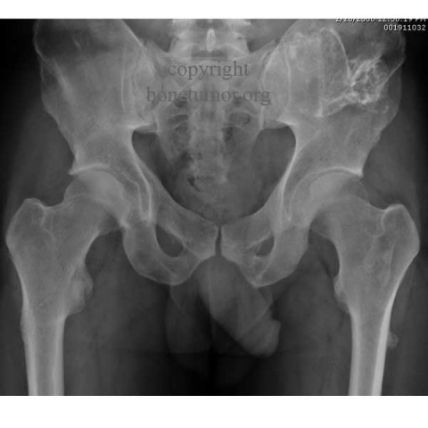
This very pleasant gentleman is 32 and he works as a mechanical engineer. He is here because of multiple hereditary exostosis (HMOCE). His grandfather, grandmother, and two brothers are affected. He has had several surgeries for removal of osteochondromas. Most importantly, a lesion has been partially removed from the posterior left hip. This bump has now been getting bigger and more painful. Now, there appears to have been change in the left hip lesion, which appears to be larger. I have recommended surgical removal of the left hip lesion. We plan wide excision left iliac bone tumor.
Radiological findings:
In the posterior left iliac crest there is one palpable lesion which is about 6 x 6 cm, and according to the patient has enlarged.This is the one that may have been changing. On the left wrist there is about 15 to 20% loss in both supination and pronation, and over radial side of the wrist on the distal radius there is a palpable osteochondroma. New and old MRI scans are also compared, there has been definite enlargement of the mass in the left posterior ilium. The extraosseous surface volume of the lesion has roughly doubled in size in six months. On plain radiographs of the left hemipelvis there is a large lesion which has been surgically treated on at least one occasion. A significant amount of abnormal bone remains. Comparison of old and new radiographs show some enlargement of the lesion in the left iliac crest. Two MRIs made several months apart document that there has been significant enlargement of the lesion, which has a large area of high signal intensity.
Differential Diagnosis
This patient has documented HMOCE ( osteochondromatosis). The radiographic findings and the patient's history as well as the family history absolutely confirm the diagnosis. The only question is what treatment should be given.
Special Features of this Case:
As you read in the material above, the diagnosis is confirmed - what is that diagnosis? What treatment would recommend?
Imagen

Case ID Number
Image Types
Image modality
Tumor Name
Tumor Type
Benign or Malignant
Body region
periosteal reaction
position within the bone
Tumor behavior
Tumor density


