Case Identification
Case ID Number
Tumor Type
Body region
Position within the bone
Benign or Malignant
Clinical case information
Case presentation
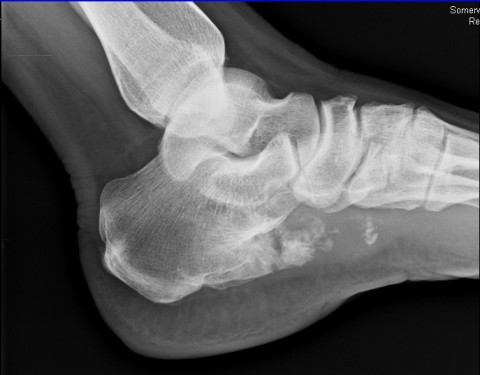
The patient reports an enlarging mass in the left foot for 2 years. Apparently it was small 2 years ago and now is quite substantial. It is painful, she cannot put on a regular shoe, and it is starting to significantly interfere with function
Radiological findings:
There is a bony mass that projects from the plantar lateral surface of the calcaneus. The mass appears to be arising from the surface of the calcaneus, almost directly plantar. The portion adjacent to the calcaneus is ossified, the more superficial and lateral portion is lucent, with scattered calcifications. It appears that the cortex of the calcaneus and the cortex of the lesion are confluent at the base of the lesion. The normal trabecular bone of the medullary portion of the calcaneus appears to continues out into the base of the lesion. There is cap of tissue covering the surface of the lesion and projecting from its lateral border, that is more than 2 cm thick in some areas, lobular, with very bright T2 signal on MRI suggestive of cartilage.
Laboratory results:
No laboratory examinations were requested.
Differential Diagnosis
This lesion has a bony base and a cartilaginous cap. It is a surface lesion. However, the cartilaginous cap is quite thick, and there has been progressive growth. Differential diagnosis includes osteochondroma, secondary chondrosarcoma.
Further Work Up Needed:
For biopsy purposes, large wedge shaped sections of the thick cartilaginous cap were excised and sent for pathologic examination.
Special Features of this Case:
Several authors have reported that benign osteochondroma can be distinguished from secondary chondrosarcoma based on the thickness of the cartilage cap. A recent report shows that cap thickness of 2 cm or greater strongly indicated secondary chondrosarcomas.
Radiology. 2010 Jun;255(3):857-65. Epub 2010 Apr 14.
Improved differentiation of benign osteochondromas from secondary chondrosarcomas with standardized measurement of cartilage cap at CT and MR imaging.
Bernard SA, Murphey MD, Flemming DJ, Kransdorf MJ.
Radiology. 2010 Jun;255(3):857-65. Epub 2010 Apr 14.
Improved differentiation of benign osteochondromas from secondary chondrosarcomas with standardized measurement of cartilage cap at CT and MR imaging.
Bernard SA, Murphey MD, Flemming DJ, Kransdorf MJ.
Imagen

Case ID Number
Image Types
Image modality
Tumor Name
Example Image
yes
Benign or Malignant
Body region
position within the bone


