Case Identification
Case ID Number
Tumor Type
Body region
Position within the bone
Benign or Malignant
Clinical case information
Case presentation
A 39 year old man had a history of pain and progressive loss of motion of the left hip and knee.
Radiological findings:
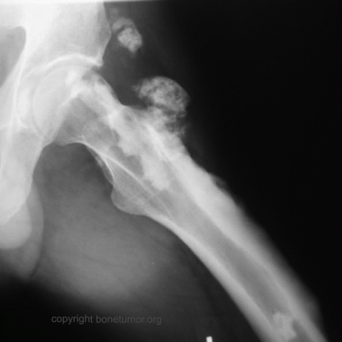
Radiographically, the lesions show undulating cortical hyperostosis which has the appearance of flowing candle wax. There are also soft tissue masses which are mineralized.
The femor lesions show narrowing of the medullary canal. The cortical hyperostosis extends across or nearly across joints and causes loss of motion.
Extensive soft tissue masses may develop, most of which are adjacent to the involved bone, but some may be unconnected to the bone. The soft tissue masses become more ossified over time. Heterogeneous signal intensity is seen on MR imaging due to the mixture of osseous, fibrous, adipose, and cartilagenous tissue these contain. The soft tissue lesions enhance with IV gadolinium. An erroneous dignosis of sarcoma is possible, particularly when the soft tisue lesion is unmineralized.
Special Features of this Case:
What is the diagnosis?
Image

Case ID Number
Image Types
Image modality
Tumor Name
Tumor Type
Benign or Malignant
Body region
Bone name
position within the bone
Tumor behavior
Tumor density


