Case Identification
Case ID Number
Tumor Type
Body region
Benign or Malignant
Clinical case information
Case presentation
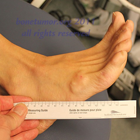
This 44 year old woman has a 3.5 by 4 cm mass on the lateral distal portion of the foot. Can a definite diagnosis be made right away in the clinic, without a complicated and expensive work up? Read on.
Radiological findings:
Plain radiographs show no abnormality other than the soft tissue swelling that can be seen clinically. This is important because bone changes or calcification in the soft tissues or in the mass would completely rule out the diagnosis that was found in this case.
Laboratory results:
no laboratory examinations are needed for this diagnosis.
Differential Diagnosis
The differential diagnosis of soft tissue masses in the foot and ankle is broad. Both benign and malignant processes must be considered. However, superficial, lobular masses that are associated with tendon sheaths or joint, that grow and shrink over time, and become more and less firm when the nearby tendon or joint is moved, may be suspected to be ganglion cyst. If history and initial physical exam point to the possible diagnosis of ganglion cyst, the diagnosis can be confirmed using the diagnostic maneuver shown in the photographs.
Further Work Up Needed:
The further workup that is required is transillumination and aspiration. To perform trans illumination, it is generally helpful to work in a low light environment. A small LED penlight or a laser pointer works best, because they create a very focused intense light that can be projected into the mass.
If transillumination is positive, the next up is aspiration. The mass is entered in one single location with a large bore needle. Multiple punctures and multiple pass of the needle are not recommended. Ganglion cyst material can be very thick and resistant to aspiration. Using a large bore needle on a small volume syringe is helpful. The presence of clear ganglion type fluid absolutely confirms the diagnosis and no further workup is needed. Aspiration of old or new blood is NOT considered a sign of a ganglion and mandates further investigation.
If transillumination is positive, the next up is aspiration. The mass is entered in one single location with a large bore needle. Multiple punctures and multiple pass of the needle are not recommended. Ganglion cyst material can be very thick and resistant to aspiration. Using a large bore needle on a small volume syringe is helpful. The presence of clear ganglion type fluid absolutely confirms the diagnosis and no further workup is needed. Aspiration of old or new blood is NOT considered a sign of a ganglion and mandates further investigation.
Treatment Options:
Positive findings on transillumination and aspiration of a suspected ganglion cyst confirms the diagnosis, and no further tumor workup is needed. In this case, recommended further treatment includes complete aspiration of as much fluid as can be removed, multiple punctures of the interior portions of the cyst wall with the same needle, and injection of cortisone. If the lesion is large or troublesome, or if the lesion recurs after the initial aspiration, surgical removal is recommended.
Special Features of this Case:
Cystic lesions that contain clear fluid will transilluminate, because the light is conducted by the fluid to the furthest extent of the mass. Normal tissues will not transilluminate in this way. Although some light passes through normal tissues, the distance the light travels is much less.
As shown in the photos, both the lesion and the nearby normal tissues are illuminated and the amount of spreading of the light is compared. Transillumination is considered positive when the entire lesion is illuminated even when the light is applied to a very peripheral portion of the mass.
As shown in the photos, both the lesion and the nearby normal tissues are illuminated and the amount of spreading of the light is compared. Transillumination is considered positive when the entire lesion is illuminated even when the light is applied to a very peripheral portion of the mass.
Image

Secret Tumor Name
Case ID Number
Image Types
Image modality
Tumor Name
Example Image
yes
Benign or Malignant


